HFpEF vs. HFrEF: How Can Heart Failure Happen With a Normal EF?
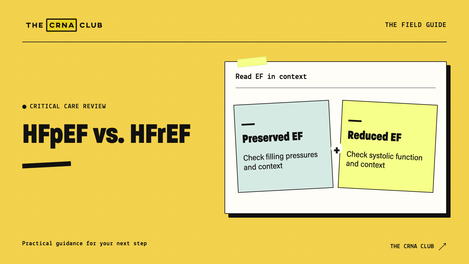
You can have heart failure with a normal ejection fraction. In HFpEF, left-ventricular filling pressures are elevated despite a preserved EF. In HFrEF, the ventricle’s pumping function and EF are reduced.
Sachi, CRNA
CRNA
In This Article (8 sections)
- What does ejection fraction actually measure?
- Which EF categories will you see in study material?
- How does impaired filling cause symptoms with a preserved EF?
- Is a normal EF enough to diagnose HFpEF?
- Does preserved EF mean less serious disease or no useful treatment?
- How would you explain HFpEF in a CRNA interview?
- Our Final Thoughts
- Keep working through cardiac cases
For CMC study and CRNA interview preparation, we suggest CMC Review and the clinical learning library. Connect the cardiac concepts to practice questions, then organize your review with the CMC study planner. Try The CRNA Club platform free for seven days.
What does ejection fraction actually measure?
Ejection fraction is the proportion of the ventricle’s end-diastolic volume ejected during systole:
EF = stroke volume ÷ end-diastolic volume × 100%.
Stroke volume is the amount ejected in a beat. End-diastolic volume is the amount in the ventricle before that contraction. A percentage describes their relationship, not the absolute amount of blood leaving the heart.
Consider two hypothetical calculations:
| Example | End-diastolic volume | Stroke volume | Calculated EF |
|---|---|---|---|
| A | 100 mL | 60 mL | 60% |
| B | 70 mL | 42 mL | 60% |
Both have the same EF, but Example B ejects less blood per beat. At a hypothetical heart rate of 80/min, their cardiac outputs would be 4.8 and 3.36 L/min respectively.
These examples explain the arithmetic. They don’t diagnose HFpEF, and they don’t mean every patient with HFpEF has a small ventricle or low resting cardiac output.
We suggest explaining that fraction out loud before memorizing categories. The CMC Review lessons and study tools help connect an echo value to filling, forward flow and the rest of the patient’s hemodynamics.
Which EF categories will you see in study material?
The 2022 AHA/ACC/HFSA framework uses these familiar categories:
- HFrEF: LVEF 40% or less.
- HFmrEF: LVEF 41% to 49%, with evidence supporting increased filling pressures.
- HFpEF: LVEF at least 50%, with evidence supporting increased filling pressures.
- HF with improved EF: a previous LVEF of 40% or less followed by an LVEF above 40%. Improvement does not establish that the original disease has disappeared.
These thresholds identify the named 2022 framework you will still encounter in teaching and clinical records. For the current HFpEF assessment, the 2026 ACC pathway uses preserved EF alongside an integrated diagnostic evaluation.
When you answer a question, name the framework you are using. More importantly, explain the physiology and the evidence of heart failure rather than treating a threshold as the diagnosis.
How does impaired filling cause symptoms with a preserved EF?
A ventricle with impaired relaxation or increased stiffness needs more pressure to accommodate filling. Pressure rises in the left ventricle and left atrium, contributing to pulmonary venous congestion. The patient develops breathlessness even though the fraction ejected looks preserved.
The problem often becomes more apparent during exertion, when flow needs to increase. Resting measurements don’t describe the whole response to exercise.
HFpEF also involves more than one isolated diastolic abnormality. Vascular changes, rhythm, right-heart function and comorbid conditions contribute to the clinical picture. Similarly, a patient with HFrEF often has impaired filling as well as reduced ejection. “Systolic versus diastolic” is an introduction, not a complete explanation of either syndrome.
For the relationship between pressure and blood volume, review preload, afterload and perfusion. A high filling pressure doesn’t automatically establish adequate forward flow or a specific ventricular volume.
Is a normal EF enough to diagnose HFpEF?
No. Breathlessness or edema plus a preserved EF needs further evaluation. Other cardiac and noncardiac conditions produce similar symptoms.
The 2026 ACC HFpEF pathway recommends integrating the clinical assessment with echocardiography and natriuretic peptides, while considering diagnostic limitations and alternative explanations.
For a case discussion, ask:
- What symptoms and examination findings support heart failure?
- What does the echo show beyond EF?
- Is there evidence of increased filling pressures, at rest or during exertion?
- What does the rhythm, valve assessment and clinical history add?
- Are lung disease, anemia or another cause contributing to the presentation?
Natriuretic peptide results also need context. A normal value doesn’t automatically exclude HFpEF, particularly in obesity. Avoid presenting one laboratory cutoff as an absolute rule-out test for every patient.
Does preserved EF mean less serious disease or no useful treatment?
No. A preserved fraction doesn’t establish mild illness, and effective treatment exists. Current management addresses congestion, appropriate disease-modifying therapy and contributing conditions.
The 2026 ACC pathway includes SGLT2 inhibitors and consideration of other therapies according to the patient’s phenotype and contraindications. This is a broader approach than treating HFpEF only with diuresis and blood-pressure control.
For study, explain what the treatment is trying to change. Relieving congestion addresses elevated filling pressures and symptoms. Other therapies aim to improve longer-term outcomes and the patient’s wider cardiovascular, kidney and metabolic health. The right combination requires the actual clinical assessment.
How would you explain HFpEF in a CRNA interview?
Practice this original prompt: “Your patient has pulmonary congestion and an EF of 60%. How is heart failure still possible?”
“EF is stroke volume divided by end-diastolic volume. A preserved fraction doesn’t establish normal filling pressures or normal cardiac reserve. In HFpEF, impaired relaxation and increased stiffness contribute to elevated filling pressures and congestion. I’d look at the clinical findings, the rest of the echo and evidence of raised filling pressures, and consider other causes rather than diagnose it from EF alone.”
Then change the question: “Does an EF improving from 30% to 55% mean the patient now has uncomplicated HFpEF?” Review the previous diagnosis and trajectory. Improved EF doesn’t establish cure or erase the need for continued assessment and appropriate therapy.
Use the Mock Interview Lab to practice explaining the reasoning after you’ve worked through a case yourself. Personalized study guides require eligible membership access, including the active platform trial.
Our Final Thoughts
When you read an EF, ask what happens during filling and what reaches the circulation. Then connect those findings to the patient’s symptoms. You’ll explain heart failure more accurately than you would by stopping at “the squeeze looks normal.”
Keep working through cardiac cases
Use CMC Review, the study planner and practice questions to organize the cardiac topics you need to revisit. Start the seven-day platform trial to explore the wider learning library and interview tools.