STEMI vs. NSTEMI: What the ECG Does and Does Not Tell You
STEMI and NSTEMI are myocardial infarction presentations classified by the ECG: whether the defining ST-elevation pattern is present. Both involve injury to heart muscle from ischemia, meaning the muscle isn’t getting enough oxygen for what it needs. NSTEMI doesn’t automatically mean a partly blocked artery or a smaller emergency.
Sachi, CRNA
CRNA
In This Article (8 sections)
As you study ACS, practice separating what each test shows from what you still need to find out. You don’t have to make the ECG answer every question.
To compare them, start with what the ECG tells you, then add what troponin tells you. The ACS and ECG lessons in the CRNA Club cardiac library through the seven-day platform trial help you practice putting those findings together.
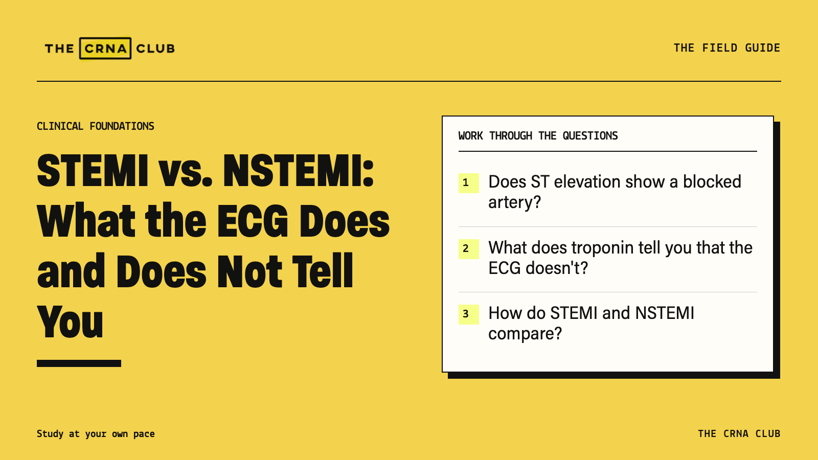
Does ST elevation show a blocked artery?
A regional ST-elevation pattern in a patient with ischemic symptoms raises concern for an acute coronary occlusion. That’s a blocked coronary artery, and the patient needs the appropriate urgent assessment and treatment pathway.
But how are you seeing the problem? Through electrical changes on the ECG. You aren’t looking inside the artery. The timing, the part of the heart affected and the leads you’re recording all change what appears. Existing electrical abnormalities can make the tracing harder to interpret too.
Some coronary occlusions don’t produce the classic pattern on a standard 12-lead. And some ST elevation has a cause other than acute coronary occlusion. That’s why you read the tracing with the patient’s symptoms and condition.
The 2025 ACS guideline guides the urgent response. If the presentation suggests an acute occlusion, don’t wait for a troponin rise before starting the indicated pathway.
What does troponin tell you that the ECG doesn’t?
Troponin gives you evidence of heart-muscle injury. A rise or fall with a value above the appropriate reference limit supports acute injury. It still doesn’t tell you why the injury happened.
Ischemia can injure the muscle, but nonischemic problems can too. So you need more than “the troponin is positive” to call the event an infarction. Look for evidence of ischemia in the symptoms, ECG and, when appropriate, imaging. Then work out the cause.
Practice that difference with the cardiac lessons. Ask what you know after each result and what you still need to find out. Our troponin explanation goes further into injury versus infarction.
How do STEMI and NSTEMI compare?
| Question | STEMI | NSTEMI |
|---|---|---|
| What does the ECG show? | A qualifying ST-elevation presentation or relevant acute-occlusion pattern | Infarction without the defining ST-elevation presentation |
| Is heart muscle injured? | Yes | Yes |
| Does the label show the exact artery anatomy? | No | No |
| Can you dismiss an occlusion because ST elevation is absent? | No | No |
| What guides urgency? | The acute pathway and the patient’s condition | Ongoing ischemia, instability and other risk findings |
The absent ST-elevation pattern doesn’t make the patient safe. It tells you which ECG presentation you’re dealing with. You still have to assess what’s happening to the patient.
How does the 2026 MI definition fit?
STEMI versus NSTEMI describes the ECG presentation. Primary, secondary and procedure-related MI describe the cause or circumstances. Those classifications answer different questions.
The Fifth Universal Definition of MI replaced the older numerical cause categories with those terms in 2026. It kept the ECG distinction. It also emphasizes troponin reference limits specific to the assay and sex, and imaging to help clarify the cause.
That means an older “type 2” label isn’t a shortcut for every troponin rise during illness. First decide whether there’s injury and evidence of ischemia. Then use the current definition to describe what caused the event.
What do two different presentations show you?
In these fictional practice examples, Patient A has ongoing ischemic symptoms and a qualifying regional ECG pattern. The urgent assessment and reperfusion pathway shouldn’t wait for troponin to rise. Reperfusion means restoring blood flow to the affected muscle.
Patient B has worsening ischemic symptoms and rising troponin, without qualifying ST elevation. That patient still needs prompt assessment. Recurrent symptoms or hemodynamic compromise increase the concern even though the ECG doesn’t show the classic pattern.
Neither case lets you name the exact angiographic finding from the monitor. Both give you enough information to recognize why waiting could be dangerous.
Which shortcuts should you stop using?
Don’t turn NSTEMI into “partial blockage,” or no ST elevation into “no emergency.” An elevated troponin alone also doesn’t confirm MI. And stable angina doesn’t belong in the acute coronary syndrome group.
Instead, ask: is there acute injury? Is there evidence of ischemia? What does the ECG show? How is the patient doing right now?
Our Final Thoughts
Choose one ACS presentation and talk through the findings before you give it a label. What does the ECG show? What does troponin show? What makes the patient urgent? Keep practicing that distinction if the labels are easier to recall than the reasoning behind them.
Continue your ACS review
Work through the ACS and ECG material in the CRNA Club cardiac library through the seven-day platform trial. Use one case to explain the evidence for injury and ischemia, then name what you still need to know.