Cardiogenic Pulmonary Edema vs. ARDS: Pressure and Permeability
Cardiogenic pulmonary edema mainly develops when high pressure in the pulmonary circulation pushes fluid into the lungs. In ARDS, inflammatory injury makes the gas-exchange barrier more permeable, so fluid leaks through it. Both impair oxygenation, and both mechanisms can be present in the same critically ill patient.
Sachi, CRNA
CRNA
In This Article (8 sections)
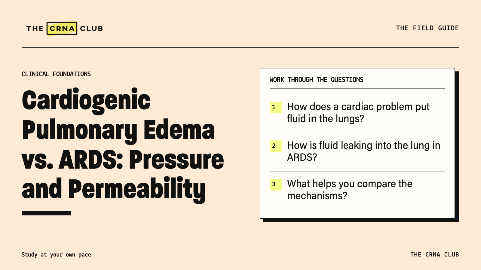
- How does a cardiac problem put fluid in the lungs?
- How is fluid leaking into the lung in ARDS?
- What helps you compare the mechanisms?
- Does the P/F ratio distinguish them?
- What if a patient has findings of both?
- Does improvement with diuresis rule out ARDS?
- Our Final Thoughts
- Continue comparing the respiratory mechanisms
As you work through respiratory cases, follow how the fluid reached the lung before getting caught up in the diagnostic labels. That gives you a starting question for both patterns.
Start by asking why the fluid left the circulation. The respiratory and cardiac lessons in the CRNA Club clinical library through the seven-day platform trial help you follow pressure or barrier injury into the oxygenation problem.
How does a cardiac problem put fluid in the lungs?
When left atrial and pulmonary venous pressures rise, pressure in the pulmonary capillaries rises too. That’s hydrostatic pressure, the pressure pushing fluid out of the vessels.
If more fluid filters out than the lung can clear, it accumulates in the tissue and alveoli. Blood still reaches poorly aerated regions, but those regions exchange gas less effectively. That produces a shunt-like component and lowers arterial oxygen.
A weak left-ventricular contraction is one possible cause. High filling pressures can also occur with preserved EF. Acute valve disease, ischemia and abrupt changes in the load on the heart can contribute too.
So a preserved EF doesn’t end the cardiac assessment. You need to know the filling pressures and what changed.
How is fluid leaking into the lung in ARDS?
Inflammation injures the barrier between the pulmonary capillary and the alveolus. It becomes more permeable, allowing protein-rich fluid into the lung.
Surfactant, which helps keep the small air spaces open, is disrupted. Alveoli become harder to keep open, the lungs become less compliant and oxygenation worsens. Less compliance means more difficulty expanding the lung for a given pressure change.
That mechanism needs a matching clinical picture. ARDS includes an appropriate acute course, a precipitating context, imaging and oxygenation findings. One low oxygen result or one image doesn’t diagnose it. The global ARDS definition describes the criteria and support conditions.
Use the respiratory lessons to follow barrier injury through alveolar flooding to impaired gas exchange. Then the diagnostic criteria describe a process you understand.
What helps you compare the mechanisms?
| Question | Mainly hydrostatic or cardiogenic edema | ARDS |
|---|---|---|
| Why is fluid moving? | Raised vascular pressure | Inflammatory injury increases permeability |
| What history helps? | A cardiac trigger or evidence of high left-sided filling pressure | A compatible acute inflammatory or lung-injury trigger |
| Does EF decide it? | No, filling pressure can be high with preserved EF | No, cardiac disease can coexist |
| Does an image settle the cause? | No | No |
| Can mechanisms overlap? | Yes | Yes |
Put the history and examination beside fluid balance, echocardiography and other available findings. You are working out why lung water increased, rather than choosing a cause from the chest image alone.
Does the P/F ratio distinguish them?
No. The P/F ratio describes arterial oxygen relative to the inspired oxygen fraction. It tells you about oxygenation under those support conditions, rather than what caused the impairment.
In this fictional example, PaO2 80 mmHg ÷ FiO2 0.80 = 100 mmHg. The oxygenation is concerning, but the calculation doesn’t tell you whether pressure, permeability or both caused it.
Our P/F-ratio guide goes through the calculation. Keep the ratio attached to the oxygen support and measurement conditions, then assess the rest of the ARDS criteria.
What if a patient has findings of both?
Compare these fictional cases. Patient A develops acute dyspnea after a cardiac trigger, with evidence of markedly elevated left-sided filling pressure. That gives you a reason for pressure-driven filtration.
Patient B has worsening oxygenation after an inflammatory insult, bilateral opacities and respiratory failure that hydrostatic edema alone doesn’t explain. Assess the complete criteria for ARDS.
Now add ventricular dysfunction and a strongly positive fluid balance to Patient B. The permeability problem hasn’t automatically disappeared. Pressure and extra fluid can add to it. Work out how much each contributes and follow the response to the treatment plan.
Does improvement with diuresis rule out ARDS?
No. Changing fluid balance can change lung water in more than one condition. Improvement adds information, but it doesn’t prove the edema was only cardiogenic.
A single filling-pressure measurement has limits too. Technique and support conditions affect it. An old wedge-pressure cutoff shouldn’t be used alone to exclude ARDS.
Our Final Thoughts
Take one pressure-driven case and one permeability case, and draw how fluid reaches the alveoli. Then let the mechanisms overlap in the second case. You can keep the comparison simple while still explaining why a single image or oxygenation number doesn’t choose the cause for you.
Continue comparing the respiratory mechanisms
Review ARDS, respiratory failure and heart failure in the seven-day CRNA Club platform trial. Explain the fluid movement first, then put the oxygenation findings back into the case.